3D Neurosurgery · a free 3D resource for brain and spine surgeryTürkçe · beyincerrahi.org

Free resource · brain and spine surgery · 3D teaching

We teach surgery in three dimensions.

A free resource for brain and spine surgery: 3D models built from real anatomy, short animations and learning objectives for every work. Open to everyone; built by measurement, checked by a surgeon. Download, use in teaching and talks, cite the source.

  • Cost None
  • Registration None
  • Licence Media CC BY-SA 4.0 · text CC BY 4.0
  • Formats MP4 · GLB · JPG
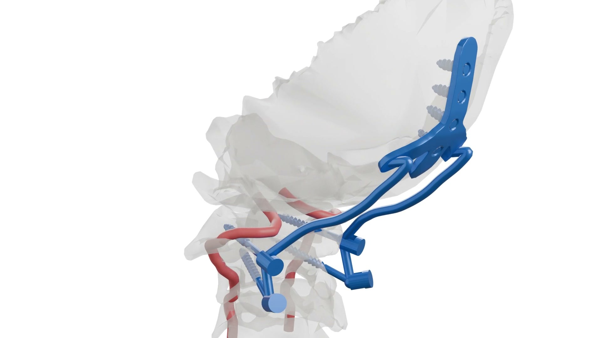
Figure 1. Plate and screw placement from the back of the skull to the second neck vertebra — 10 seconds from the team’s own animation (silent). Every screw was positioned by measurement on the model. Full animation · 50 s

The resource is growing. Every new 3D work is added here together with its learning objectives. No registration, no fee.

What we do

We model
From open anatomy data, by measurement. No line is drawn by eye.
We explain
With 20–50 second animations: entry point, angle, depth, the neighbouring vessel and nerve.
We measure
Every distance and angle is measured on the model and compared with published values; a surgeon corrects what is off.
We share
Free and open to everyone: models and videos under an open licence, for use in teaching and talks.

Turn the model yourself

The scene from the animation: the back of the skull (occipital bone), the first three neck vertebrae, the vertebral arteries, the spinal cord and the implants. Drag to rotate, scroll to zoom.

Loading the 3D model…

The same model is used in the team’s 3D anatomy atlas (3d.drozguraksan.com). Bone and vessel models come from the open-source Z-Anatomy; the implants are the team’s own.

BodyParts3D - The Database Center for Life Science - CC-BY-SA 2.1 Japan Z-Anatomy - The libre 3D atlas of anatomy - CC-BY-SA 4.0

3D works

For each work: what it shows, what it teaches, which files exist. Objectives are written and verified by a surgeon.

Animation · Craniocervical junction · 50 s

Occipitocervical fixation

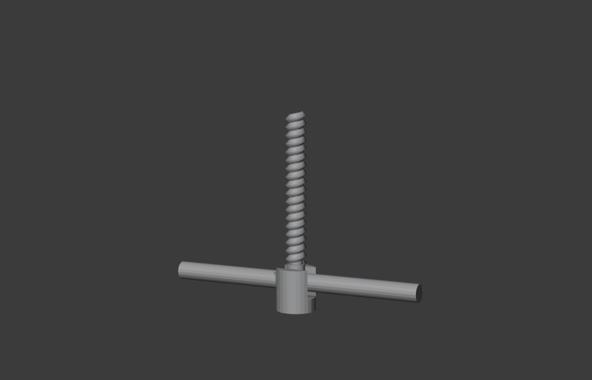
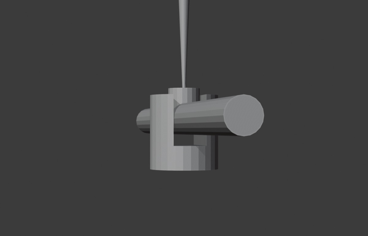
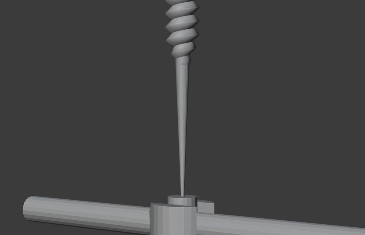
Plate, screw and rod placement from the back of the skull (occiput) to the second neck vertebra; eight stages in their real order. Screws turn with their real thread pitch.

Learning objectives
  1. Lists the stages of occiput–C2 fixation in order: entry, pilot hole, probing, tap, screw, plate, rod, O–C2 angle.
  2. Describes the entry point and trajectory of the C1 and C2 screws.
  3. Explains why the distance between screw and vertebral artery is measured.
  4. Distinguishes the parts of a polyaxial screw: threaded shaft, smooth neck, spherical head, tulip, rod.
Measurements

Measured on the model: C1 screw → vertebral artery 7.04 mm · C2 screw → vertebral artery 4.22 mm · foramen magnum sagittal diameter 37.0 mm.

Interactive 3D model · Craniocervical junction

Skull base, C1–C3, vertebral arteries and implants

The very scene from the animation. Drag, zoom; see the path of each screw inside the bone from every angle.

Learning objectives
  1. Recognises the occipital bone, C1, C2 and C3 in three dimensions; shows that the atlas has no body.
  2. Follows the course of the vertebral artery around C2 and C1.
  3. Examines the relation of the implants (occipital plate, C1 and C2 screws, rods) to the bone from every angle.
Measurements

Bone and vessels from Z-Anatomy; implants are the team’s own model. Derived from a single individual.

Interactive atlas · 188 structures · 11 models

3D anatomy atlas

Lumbar, cervical and thoracic spine, skull and cranial nerves, craniocervical junction, a spondylolisthesis scenario. Sectioning, exploded view, layer toggles; every structure explained in Turkish and English.

Learning objectives
  1. Names the bone, disc, dura and nerve root at every level of the spine.
  2. Matches the exit levels of the nerve roots with their dermatome bands.
  3. Shows how the canal narrows in spondylolisthesis.
  4. Compares the position of a pedicle screw inside the vertebra with the bone transparent and opaque.
Measurements

Spinal cord, root, cranial nerve and vertebral artery diameters were compared with MRI and cadaver series and corrected.

In preparation

Cervical: unilateral approach for bilateral decompression
Laminotomy, contralateral undercutting, removal of the flavum, cord decompressed. In production.
Lumbar: epidural, root, facet and SI joint injections
One needle path in two windows: 3D anatomy and virtual fluoroscopy (AP · lateral · oblique). In production.
C1 lateral mass and C2 pedicle screws
Entry points, trajectories and the vertebral artery corridor, by measurement. Planned.

How the learning objectives are written

Three to four objectives per work. An objective is what the viewer can do at the end; it is written with a measurable verb.

Verb
Lists, describes, distinguishes, shows, explains. No “knows” or “understands”.
Scope
One work, one scene. An animation teaches one step of an operation, not the whole operation.
Verification
A surgeon writes the objective; if there is a number, it is measured on the model and placed next to the published value.
Version
When the work changes, the objective is rewritten; the old version is not deleted.

Why this way

Three reasons. The numbered evidence is below; references link to free sources.

3D teaches anatomy better
That is the pooled result of dozens of studies, and the gap widens when the model is interactive. A young surgeon trained on a simulator places screws more accurately. [1] [2] [5]
Anatomy differs from person to person, so we measure
When a screw in the second neck vertebra is planned on CT, the vertebral artery is at risk in one of three patients; the same level has four different groove types. Building a model by eye is not enough. [9] [10]
AI often gets anatomy wrong; the surgeon checks
Most anatomy drawings from text-to-image tools come out wrong. We do not let AI draw anatomy: it builds the scene, and we measure the result. [17] [18]
This kind of training has not been measured yet
We found no study evaluating training in which surgeons produce their own 3D teaching material (our two searches: 716 and 1,264 records). The closest study used a model made by the teacher. The objectives in this resource are written for that gap. [8]
Evidence — 18 studies, with their numbers

3D teaches

Moderate gain in spatial knowledge
3D visualisation improves spatial knowledge by d = 0.50 and factual knowledge by d = 0.30 compared with other methods (36 studies, 2,226 students). [1]
Interaction is essential
Interactive stereoscopic 3D outperforms interactive monoscopic 3D (ES = 0.53); passively viewed stereo shows no added effect (13 RCTs). [2]
Strongest as a complementary resource
Virtual and augmented reality used as a complementary resource: SMD = 0.52; versus lectures alone: 1.00 (27 studies, 2,199 students). [3]
Screw penetration from 30% to 7%
Force-feedback virtual training reduced thoracic pedicle screw penetration from 30% to 7.14% (randomized cadaver study, 10 residents). [4]
More accurate screws in real patients
Junior surgeons trained on an immersive VR simulator achieved pedicle screw success of 82.9% vs 74.2% (double-blind RCT, 24 surgeons). [5]
A free simulator halves the error
After two sessions on an open-access web simulator, screw misplacement fell from 50% to 20.8% (RCT, 24 students). [6]
Patient-specific 3D model with mixed reality
Exam scores: ventricular puncture 20.25 vs 17.38, hematoma puncture 20.75 vs 17.13 (P < 0.01; 16 physicians, randomized). [7]

Anatomy varies, measurement carries error

Artery at risk in one of three C2 patients
C2 pedicle screw simulation in 280 patients: vertebral artery injury predicted in 35% (98/280); narrow pedicle (OR 3.49) and high-riding artery (OR 4.74) were independent risk factors. [9]
Same level, four different grooves
The C2 vertebral artery groove fell into four types across 90 sides (58.9 · 17.8 · 14.4 · 8.9%); one type required a translaminar rather than a pedicle screw. [10]
Not everyone has a C1 pedicle
Of 84 atlas pedicles (Indian population), about 32% were thinner than 4 mm and 38% had no pedicle; significantly thinner in women. [11]
Planned entry point: breach from 32% to 14%
In 300 C2 pedicles, a 5-mm cylinder breached 32% from the standard entry point and 14% when planned to the isthmus. [12]
Cobb angle: ±7.5°
Repeat measurements by the same observer differed by a mean of 2.6°; between observers, a single measurement had a 95% confidence interval of ±7.5° (CT reformats, major curves). [13]
Viewing at an angle adds 10°
When the radiograph was photographed at 45° and measured, the mean Cobb angle rose from 35.1° to 45.0° (P < 0.001). [14]
Digital alone is not enough
Digital tools did not improve accuracy by themselves; inter-observer ICC for end-vertebra selection was 0.83 manually and 0.74 digitally. [15]
Right tool, right method
Smartphone-assisted end-vertebra selection reduced the 95% error limit of the Cobb measurement from 3.6° to 1.9°; inter-observer ICC rose from 0.910 to 0.967. [16]

AI gets it wrong

80.8% anatomically incorrect
In an expert rating by 33 reviewers of 110 DALL·E 3 images of congenital heart disease, 80.8% were anatomically incorrect or fabricated and 85.2% had incorrect labels. [17]
37.4% fully correct in the best model
Six generators, 144 images: the share of fully correct anatomical structures was 37.4% in the best model, lower for complex structures. [18]
Errors reaching print
In 2025, 10.8% (4 of 37) of aesthetic medicine articles depicting facial anatomy contained AI images; all four showed gross anatomical errors. [19]

This course does not reject AI. It teaches you to give it the right data and check the result by measurement.

How to use

In lectures
Download the video and put it on a slide; the licence lines appear in the last four seconds — do not cut them.
In talks
Link to the 3D model with a QR code; the audience turns it on their phones.
With attribution
Cite the source every time (ready-made line below). If you modify and share, share under the same licence.
Not for patients
This page is for surgeons; it contains no patient information or appointments.

Dedication · Şerefeddin Sabuncuoğlu

Amasya · c. 1385 – after 1468 [20]

A miniature from Cerrâhiyyetü’l-Hâniyye: a physician applies a cautery to the head of a seated patient
Figure 2. A miniature from Cerrâhiyyetü’l-Hâniyye: the physician applies cauterisation to the patient’s head. Public-domain scan — Wikimedia Commons, R212F1.jpeg.

His Cerrâhiyyetü’l-Hâniyye (1465) was the first Turkish surgical text to depict operations in coloured miniatures. From skull fractures and hydrocephalus to spinal injuries and sciatica, he turned surgical knowledge into images. Five and a half centuries later, teaching neurosurgery in three dimensions, we follow his path.

Licence and attribution

Anatomy
Anatomy meshes derive from Z-Anatomy (CC BY-SA 4.0), which is based on BodyParts3D (© DBCLS, CC BY-SA 2.1 JP). The models were modified: simplified, merged, implants and nerve roots added.
Team’s own work
Nerve root and implant models are the team’s own and are shared under the same licence; they do not represent any commercial product.
Media
3D models, animations, videos and images: CC BY-SA 4.0 — credit the source; if you modify and share, share under the same licence.
Text
Page text and learning objectives: CC BY 4.0.
Patient data
No patient image or personal health data was used; bone models derive from the single anonymous individual of BodyParts3D.
Notice
For education; not a clinical decision tool.
Names
The names “3D Neurosurgery”, “beyincerrahi.org” and “openbvs” are not covered by the licence.
Inside the files
The licence text is also written into the files: GLB (asset.copyright), MP4 (copyright tag) and the card in the last four seconds of the video.
Version
Animation v04.1 · loop v01.1 · 3D model v03 · poster v01.1 — 8 October 2026. Older versions are not deleted; they stay in the archive.

BodyParts3D - The Database Center for Life Science - CC-BY-SA 2.1 Japan Z-Anatomy - The libre 3D atlas of anatomy - CC-BY-SA 4.0

Z-Anatomy · BodyParts3D (DBCLS) · CC BY-SA 4.0 · CC BY-SA 2.1 JP · CC BY 4.0

Ready-made citation

3D Neurosurgery / beyincerrahi.org (openbvs). Occipitocervical fixation — 3D animation, version v04.1 (2026). CC BY-SA 4.0. https://3dneurosurgery.org/media/ccj-fiksasyon-anim-50sn-v04.1.mp4

References

Linked to the free full text (PMC) where one exists, otherwise to the PubMed record; the DOI goes to the publisher.

  1. Yammine K, Violato C. A meta-analysis of the educational effectiveness of three-dimensional visualization technologies in teaching anatomy. Anat Sci Educ. 2015. PubMeddoi:10.1002/ase.1510
  2. Bogomolova K, et al. Stereoscopic three-dimensional visualisation technology in anatomy learning: a meta-analysis. Med Educ. 2021. Free full text (PMC)PubMeddoi:10.1111/medu.14352
  3. García-Robles P, et al. Immersive virtual reality and augmented reality in anatomy education: a systematic review and meta-analysis. Anat Sci Educ. 2024. PubMeddoi:10.1002/ase.2397
  4. Hou Y, et al. Effectiveness of the thoracic pedicle screw placement using the virtual surgical training system: a cadaver study. Oper Neurosurg. 2018. PubMeddoi:10.1093/ons/opy030
  5. Xin B, et al. The efficacy of immersive virtual reality surgical simulator training for pedicle screw placement: a randomized double-blind controlled trial. Int Orthop. 2020. PubMeddoi:10.1007/s00264-020-04488-y
  6. Chatelain LS, et al. Pedagogy in spine surgery: developing a free and open-access virtual simulator for lumbar pedicle screws placement. Eur Spine J. 2023. PubMeddoi:10.1007/s00586-022-07501-7
  7. Peng Y, et al. Application effect of head-mounted mixed reality device combined with 3D printing model in neurosurgery ventricular and hematoma puncture training. BMC Med Educ. 2023. Free full text (PMC)PubMeddoi:10.1186/s12909-023-04659-6
  8. Mellal A, et al. Evaluating the impact of a hand-crafted 3D-printed head model and virtual reality in skull base surgery training. Brain Spine. 2025. Free full text (PMC)PubMeddoi:10.1016/j.bas.2024.104163
  9. Ye B, et al. Development and validation of machine learning models to predict vertebral artery injury by C2 pedicle screws. Eur Spine J. 2025. PubMeddoi:10.1007/s00586-025-09168-2
  10. Wang J, et al. An anatomic consideration of C2 vertebrae artery groove variation for individual screw implantation in axis. Eur Spine J. 2013. Free full text (PMC)PubMeddoi:10.1007/s00586-013-2779-4
  11. Srivastava A, et al. Morphometric study of C1 pedicle and feasibility evaluation of C1 pedicle screw placement with a novel clinically relevant radiological classification in an Indian population. Asian Spine J. 2017. Free full text (PMC)PubMeddoi:10.4184/asj.2017.11.5.679
  12. Davidson CT, et al. Planning C2 pedicle screw placement with multiplanar reformatted cervical spine computed tomography. J Craniovertebr Junction Spine. 2019. Free full text (PMC)PubMeddoi:10.4103/jcvjs.JCVJS_116_18
  13. Adam CJ, et al. Variability in Cobb angle measurements using reformatted computerized tomography scans. Spine. 2005. PubMeddoi:10.1097/01.brs.0000169449.68870.f8
  14. Ricart PA, et al. Validity of Cobb angle measurements using digitally photographed radiographs. Spine J. 2011. PubMeddoi:10.1016/j.spinee.2011.09.003
  15. Gstoettner M, et al. Inter- and intraobserver reliability assessment of the Cobb angle: manual versus digital measurement tools. Eur Spine J. 2007. Free full text (PMC)PubMeddoi:10.1007/s00586-007-0401-3
  16. Pepe M, et al. Use of the smartphone for end vertebra selection in scoliosis. Acta Orthop Traumatol Turc. 2017. Free full text (PMC)PubMeddoi:10.1016/j.aott.2016.12.006
  17. Temsah MH, et al. Art or artifact: evaluating the accuracy, appeal, and educational value of AI-generated imagery in DALL·E 3 for illustrating congenital heart diseases. J Med Syst. 2024. PubMeddoi:10.1007/s10916-024-02072-0
  18. Strugała A, et al. Evaluation of generative artificial intelligence models in producing anatomically accurate illustrations: a comparative study of text-to-image generators. Clin Anat. 2026. PubMeddoi:10.1002/ca.70189
  19. Zhang Y, et al. AI inaccurate anatomy: prevalence of artificial intelligence-generated illustration errors in peer-reviewed aesthetic medicine publications. Plast Reconstr Surg Glob Open. 2026. Free full text (PMC)PubMeddoi:10.1097/GOX.0000000000007676
  20. Uzel İ, ed. Cerrâhiyyetü’l-Hâniyye. Ankara: Türk Tarih Kurumu; 1992. LCCN 94964112